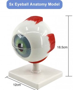
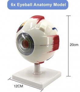
Medical Science Human Anatomical Simulation Eyeball Structural eye anatomical model 3 times larger 6 part eye anatomy model
Medical Science Human Anatomical Simulation Eyeball Structural eye anatomical model 3 times larger 6 part eye anatomy model
|
Product name
|
3 Times Enlarged Eyeball Model with mark
|
|
Size
|
12*11*20 cm
|
|
Weight
|
0.3 kgs
|
|
Color
|
Realistic shape and bright color. The model adopts Computer color matching, excellent color drawing, which is not easy to fall off, clear and easy to read, easy to observe and learn.
|
|
Packing
|
40pcs/carton, 47*26*58.5cmcm, 9kgs
|


1. CORNEA 7. VITREOUS BODY
3. CHOROID 9. FOVEA CENTRALIS
4. RETINA 10. VORTICOSE VEINS
5. IRIS 11. CILIARY MUSCLES
6. LENS 12. CENTRAL RETINAL ARTERIES AND VEINS
Human anatomy model mainly studies the systematic anatomy part of gross anatomy. The above terms in medicine come from anatomy, which is closely related to physiology, pathology, pharmacology, pathogenic microbiology and other basic medicine as well as most clinical medicine. It is the foundation of the foundation and an important medical core course. Anatomy is a highly practical course. Through the study of practice and the training of skill operation, students can enhance their ability to observe problems, solve problems, practice and think independently, and lay the foundation for future clinical operation, nursing operation and other professional skills. Anatomy is one of the examination contents of medical students’ qualification. Learning anatomy well will lay a foundation for medical students to pass these examinations successfully.


