Medical science enteric villi tissue model anatomical section of middle school student physical education instrument model
Medical science enteric villi tissue model anatomical section of middle school student physical education instrument model


Medical science enteric villi tissue model anatomical section of middle school student physical education instrument model
|
Product name
|
Human intestinal villi anatomical model
|
|||
|
Size
|
20*15*16.5cm
|
|||
|
Packing
|
20.5*17.5*17cm
|
|||
|
Usage
|
Anatomical Demonstration
|
|||

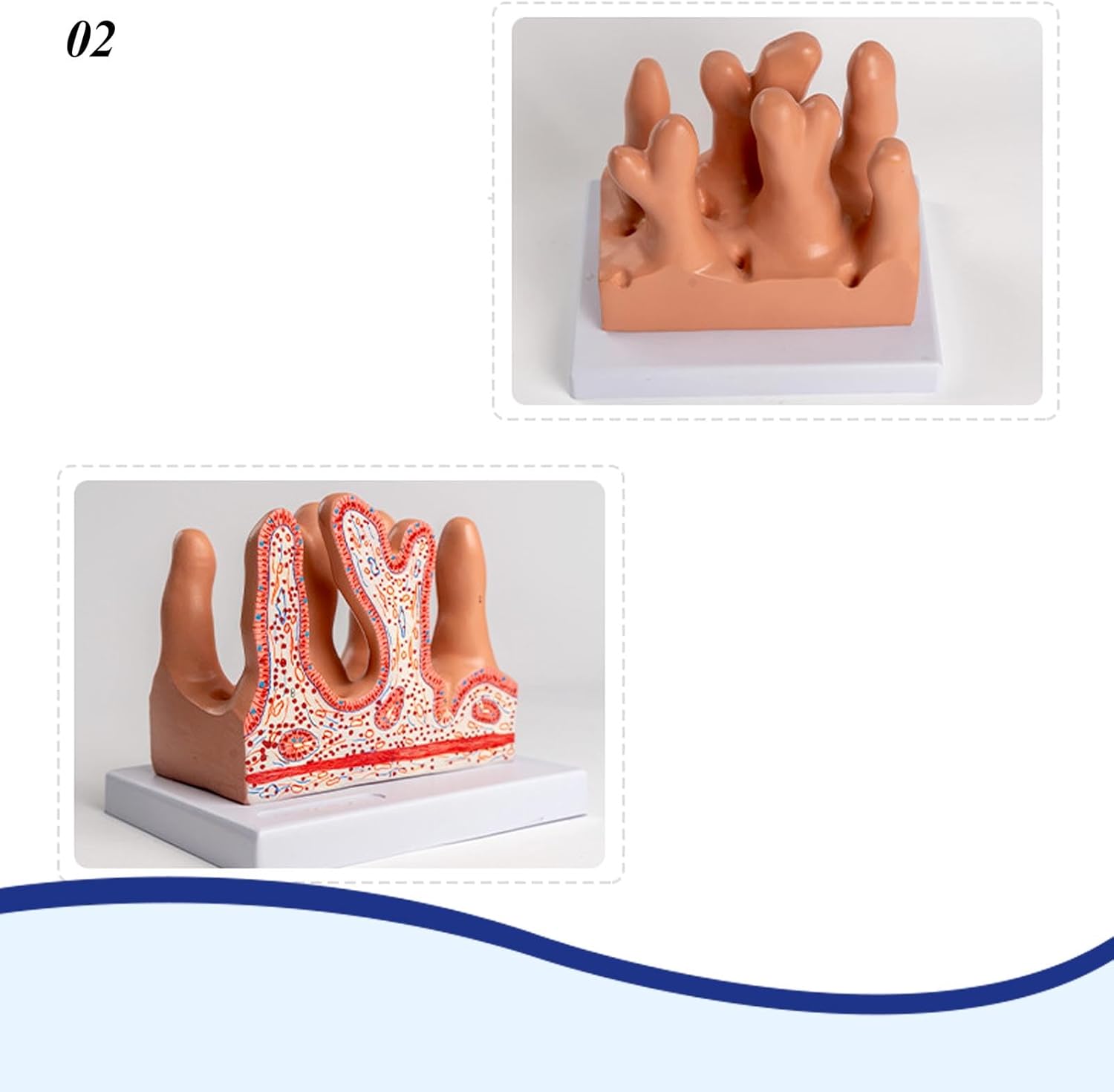
*Human intestinal villi model: This model shows that there are many villi structures on the mucosal surface of the inner surface of the jejunum. The villi are formed by the mucosal epithelium and the lamina propria that protrude into the intestinal lumen. Magnified 400 times, the anatomical structure of the intestinal villi is shown under the microscope.
*Application: Suitable for those who are interested in anatomy, nursing, physiology. Learn about the organs of the human body and their relative positions. Great for children’s education, it’s a perfect gift for kids.
*Material: The model is made of high-quality PVC material, computer color matching, high-end hand-painted, very realistic, almost realistically depicting the structure of human organs
*Digital Signage: Models We have specially designed 13 digital signages for you, so that you can learn accurately and effectively, understand and practice better. Save learning time and improve learning efficiency
*Service: If you have any questions about our products, please contact us in time, we will reply you as soon as possible within 24 hours. happy shopping!
Product Details

The model showed many villi structures on the mucosal surface of the inner jejunum. The villi were formed by the mucosal epithelium and lamina propria protruding into the intestinal lumen.


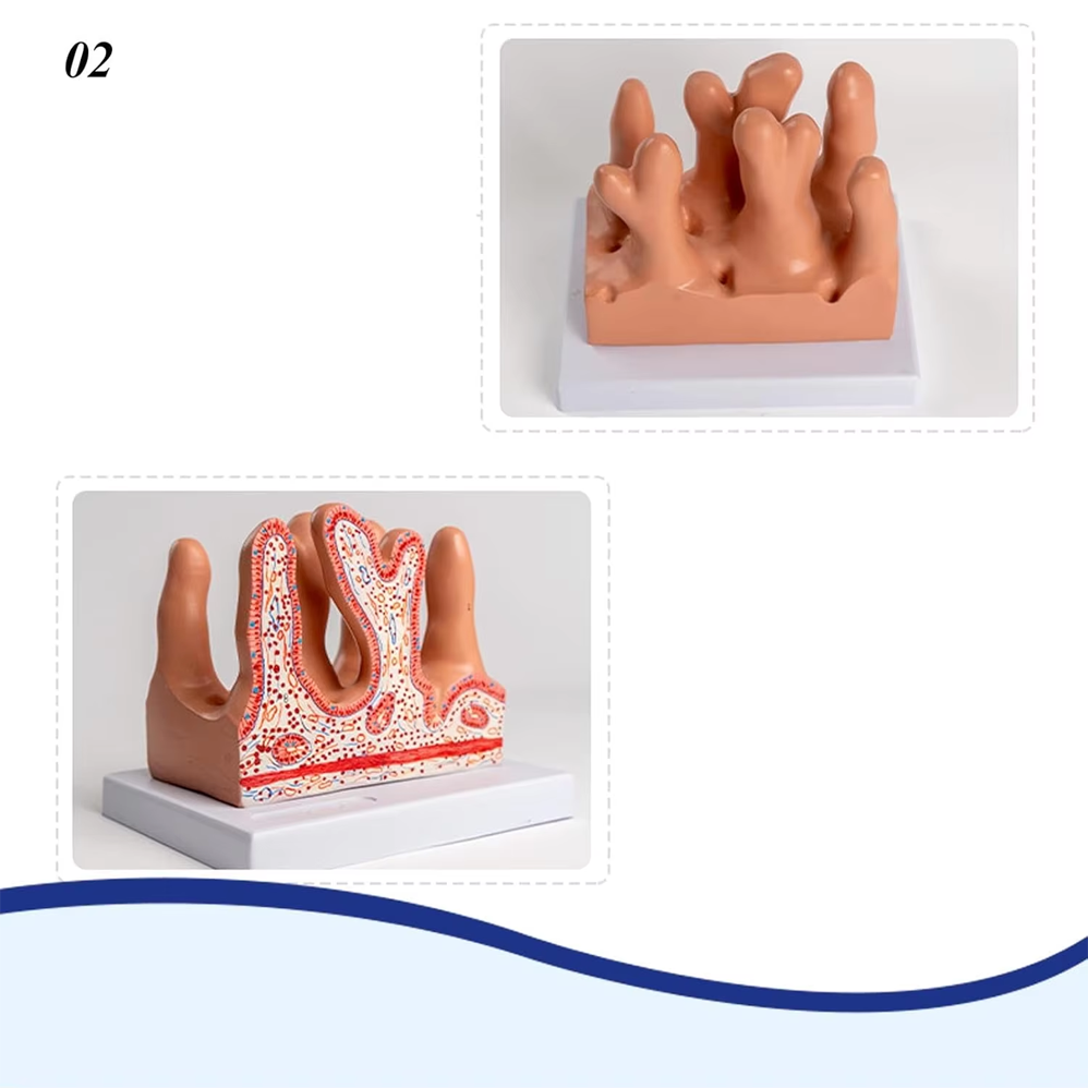
his anatomy model shows the basic anatomical detail of intestinal villus,helping to understand. Our anatomical intestinal villus
model is life-sized,perfect for digestive system diseases study, research,can be used as a teaching aid for understanding The
inner surface of the wall of the small intestine has a lot of ring-shaped folds, and there are many hairy lumps in the folds
called villi of the small intestine. Made from PVC material,resistance, lightweight design,and high strength.