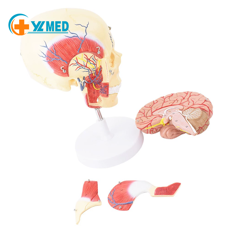
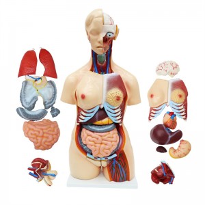
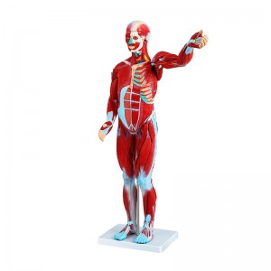
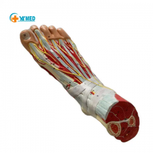
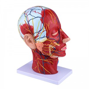
Human anatomical model of the maxillofacial anatomy of masticatory muscles Masseter temporalis trigeminal nerve
Human anatomical model of the maxillofacial anatomy of masticatory muscles Masseter temporalis trigeminal nerve
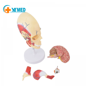
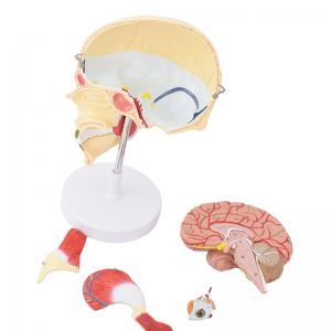
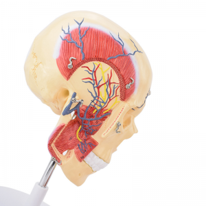
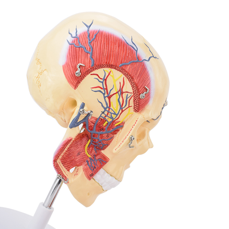
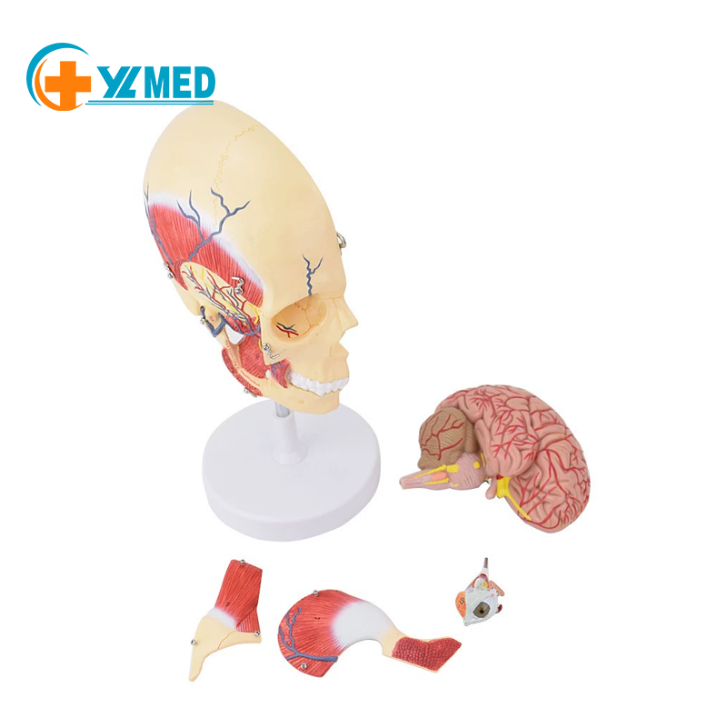
This model is designed with life-size, made of high-quality environmentally friendly PVC materials, restores the real skull brain structure, and displays the anatomical structure of the brain and other highly accurate. The maxillary muscle group and the lower jaw muscle group can be beaten, and you can see the deep nerve arteriovenous and muscle anatomy of the maxillofacial region.
1.Product Material
High quality and environmentally friendly PVC. The PVC raw materials are non-toxic and non-polluting and can be stored for a long time.
2.Research Carefully.
Each medical model is carefully guided by experts and is fully ergonomic.
3.Painted Carefully.
According to the characteristics of the model, we choose the correct color and draw a stroke.


